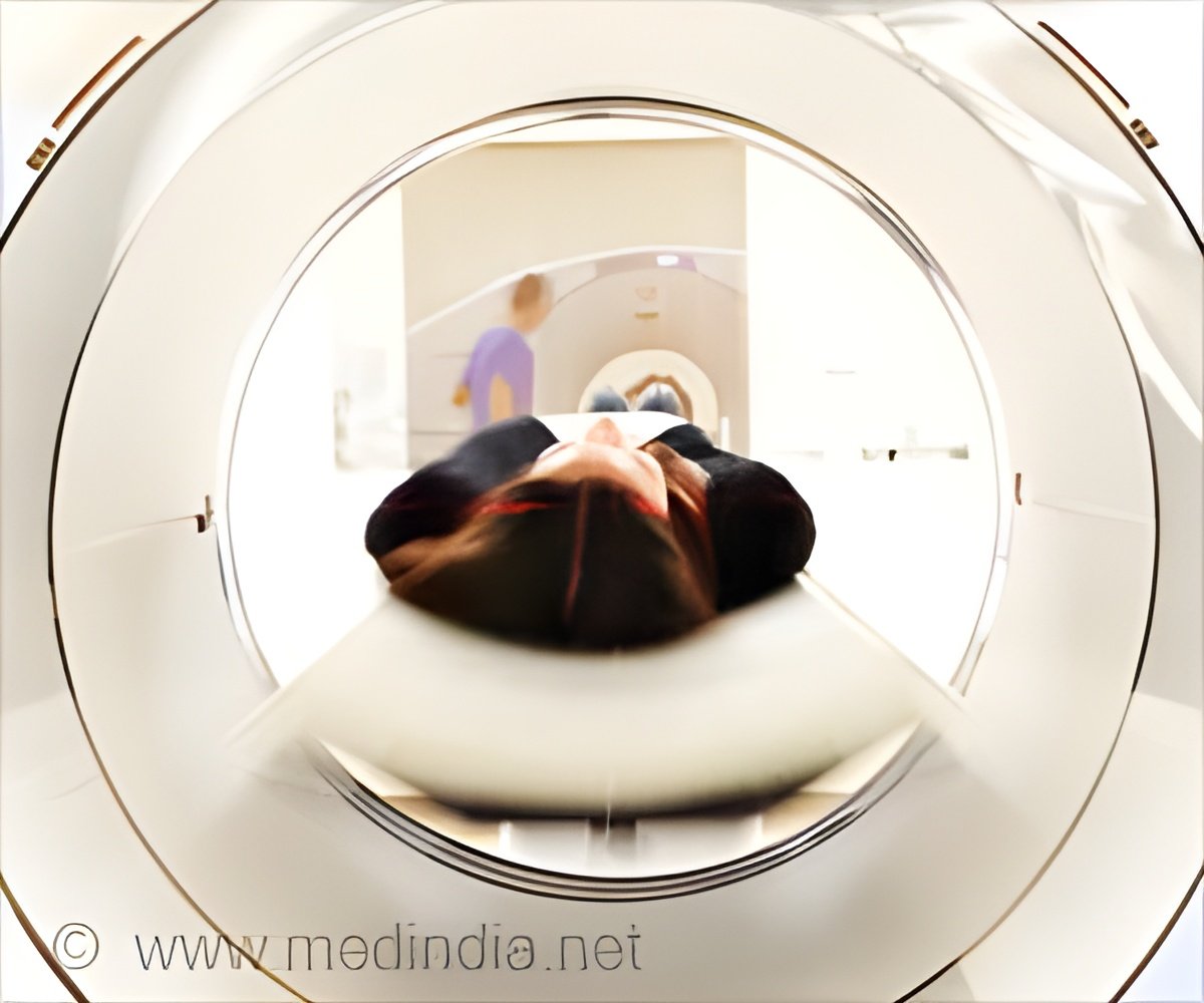
New MRI technique predicts ovarian cancer response to treatment within 48 hours, offering faster, personalized care.

Hyperpolarised carbon-13 imaging technique used in MRI scans can enhance the signal strength 10,000 times stronger providing clear images (1✔ ✔Trusted Source
Metabolic imaging distinguishes ovarian cancer subtypes and detects their early and variable responses to treatment
).
This MRI-based imaging technique developed at the University of Cambridge, predicts the response of ovarian cancer tumors to treatment and instantly shows how well the treatment works in patient-derived cell models.
Advertisement
MRI Technique Predicts and Tracks Ovarian Cancer Treatment
The technique shows whether a tumor is sensitive or resistant to Carboplatin. Carboplatin is a standard chemotherapy treatment for ovarian cancer. It will enable oncologists to predict how well a patient will respond to treatment and to see how well the treatment is working within the first 48 hours.
Different forms of ovarian cancer respond differently to drug treatments. With current tests, patients typically wait for weeks or months to find out whether their cancer is responding to treatment. The rapid feedback this new technique provides will help oncologists adjust and personalize treatment for each patient within days.
The study compared the hyperpolarised imaging technique with results from Positron Emission Tomography (PET) scans, which are already widely used in clinical practice. The results show that PET did not pick up the metabolic differences between different tumor subtypes, so could not predict the type of tumor present. The report is published today in the journal Oncogene.
“This technique tells us how aggressive an ovarian cancer tumor is, and could allow doctors to assess multiple tumors in a patient to give a more holistic assessment of disease prognosis so the most appropriate treatment can be selected,” said Professor Kevin Brindle at the University of Cambridge’s Department of Biochemistry, senior author of the report.
Advertisement
Non-Invasive Imaging Changes Ovarian Cancer Care
Ovarian cancer patients often have multiple tumors spread throughout their abdomen. It isn’t possible to take biopsies of all of them, and they may be of different subtypes that respond differently to treatment. MRI is non-invasive, and the hyperpolarised imaging technique will allow oncologists to look at all the tumors at once.
Brindle added: “We can image a tumor pre-treatment to predict how likely it is to respond, and then we can image again immediately after treatment to confirm whether it has indeed responded. This will help doctors to select the most appropriate treatment for each patient and adjust this as necessary.
“One of the questions cancer patients ask most often is whether their treatment is working. If oncologists can speed their patients onto the best treatment, then it’s clearly of benefit.” The next step is to trial the technique in ovarian cancer patients, which the scientists anticipate within the next few years.
Advertisement
Hyperpolarized Imaging Aids Ovarian Cancer Diagnosis
Hyperpolarised carbon-13 imaging uses an injectable solution containing a ‘labeled’ form of the naturally occurring molecule pyruvate. The pyruvate enters the cells of the body, and the scan shows the rate at which it is broken down – or metabolized – into a molecule called lactate. The rate of this metabolism reveals the tumor subtype and thus its sensitivity to treatment.
This study adds to the evidence for the value of the hyperpolarised carbon-13 imaging technique for wider clinical use. Brindle, who also works at the Cancer Research UK Cambridge Institute, has been developing this imaging technique to investigate different cancers for the last two decades, including breast, prostate, and glioblastoma – a common and aggressive type of brain tumor. Glioblastoma also shows different subtypes that vary in their metabolism, which can be imaged to predict their response to treatment. The first clinical study in Cambridge, which was published in 2020, was in breast cancer patients.
Each year about 7,500 women in the UK are diagnosed with ovarian cancer – around 5,000 of these will have the most aggressive form of the disease, called high-grade serous ovarian cancer (HGSOC).
The cure rate for all forms of ovarian cancer is very low and currently only 43% of women in England survive five years beyond diagnosis. Symptoms can easily be missed, allowing the disease to spread before a woman is diagnosed – and this makes imaging and treatment challenging.
Reference:
- Metabolic imaging distinguishes ovarian cancer subtypes and detects their early and variable responses to treatment – (https://www.nature.com/articles/s41388-024-03231-w)
Source-Eurekalert


