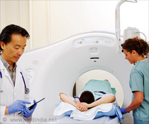
A traumatic brain injury results from a violent blow to the head. It may damage the brain thereby resulting in loss of functions and disability. Even milder forms of head injuries may have lasting damages on brain.
Hence it is necessary to establish the understanding of the short- and long-term effects of head injury for better treatments. The present study was a part of the Transforming Research and Clinical Knowledge in Traumatic Brain Injury (TRACK-TBI) study.
The team had enrolled 1,935 participants with mild TBI who underwent CT scans and were followed up till12 months after injury. Three distinct sets of patterns were identified on the CT scans. This indicated different types of damages after head injury with varying outcomes.
Brain Imaging in Head Injuries
It was found that certain types of head injuries such as contusion (bleeding into brain tissue), subarachnoid hemorrhage (bleeding into the cerebrospinal fluid over the brain), subdural hematoma (bleeding between the brain and the thick covering over the brain), and intraventricular hemorrhage (bleeding into the fluid filled spaces in the center of the brain) were associated with worse outcomes 12 months after injury.
‘Imaging after mild traumatic brain injury (TBI) may show the presence of certain features on CT scans that would help predict treatment and outcomes. This may help in further understanding the effects of head injury on brain structure and function.
’
Epidural hematoma (EDH) is a type of head injury where the bleeding lies between the skull and outer brain covering known as the dura mater. EDH was associated with incomplete recovery at two weeks and three months. However, it was not linked to negative longer-term outcomes.
The TRACK-TBI study validated with the Collaborative European NeuroTrauma Effectiveness Research in Traumatic Brain Injury (CENTER-TBI) study of 2594 TBI subjects that showed similar results. However further research is required to understand the effects of head injury on brain structure and function.
Source: Medindia


