In particular, targeting cells responsible for pulmonary fibrosis early on could possibly prevent or ameliorate long-term complications in survivors of severe COVID-19,” says Benjamin Izar, MD, PhD, assistant professor of medicine, who led a group of more than 40 investigators to complete in several months a series of analyses that usually takes years.
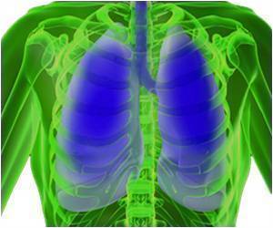
The study directly examines lung tissue using single-cell molecular profiling that can identify each cell in a tissue sample and record each cell’s activity, resulting in an atlas of cells in COVID lung.
“A normal lung will have many of the same cells we find in COVID, but in different proportions and different activation states,” Izar says. “In order to understand how COVID-19 is different compared to both control lungs and other forms of infectious pneumonias, we needed to look at thousands of cells, one by one.”
Researchers examined the lungs of 19 individuals who died of COVID-19 and underwent rapid autopsy during which lung and other tissues were collected and immediately frozen–and the lungs of non-COVID-19 patients. Researchers also compared their findings to lungs of patients with other respiratory illnesses.
Compared to normal lungs, lungs from the COVID patients were filled with immune cells called macrophages, the study found.
“In COVID-19, we see expansion and uncontrolled activation of macrophages, including alveolar macrophages and monocyte-derived macrophages,” Izar says. “They are completely out of balance and allow inflammation to ramp up unchecked. This results in a vicious cycle where more immune cells come in causing even more inflammation, which ultimately damages the lung tissue.”
“Unlike other cytokines such as IL-6, which appears to be universally prevalent in various pneumonias, IL-1beta production in macrophages is more pronounced in COVID-19 compared to other viral or bacterial lung infections,” Izar says. “That’s important because drugs exist that tamp down the effects of IL-1beta.”
The study also found that not only does SARS-CoV-2 virus destroy alveolar epithelial cells important for gas exchange, the ensuing inflammation also impairs the ability of the remaining cells to regenerate the damaged lung.
Though the lung still contains cells that can do the repairs, inflammation permanently traps these cells in an intermediate cell state and leaves them unable to complete the last steps of differentiation needed for replacement of mature lung epithelium.
“Among others, IL-1b appears to be a culprit in inducing and maintaining this intermediate cell state,” says Izar, “thereby linking inflammation and impaired lung regeneration in COVID-19. This suggests that in addition to reducing inflammation, targeting IL-1beta may help take the brakes off cells required for lung repair.”
The researchers also found a large number of pathological fibroblasts, that create rapid scarring in COVID-19 lungs. When the fibroblast cells fill the lung with scar tissue, a process called fibrosis, the lung has less space for cells involved in gas exchange and is permanently damaged.
Izar’s team closely analyzed the cells to uncover potential drug targets. An algorithm called VIPER, developed previously by Andrea Califano, Dr, chair of systems biology at Columbia University Vagelos College of Physicians and Surgeons, identified several molecules in the cells that play an important role and could be targeted by existing drugs.
“This analysis predicted that inhibition of STAT signaling could alleviate some of the deleterious effects caused by pathological fibroblasts,” Izar says.
“Our hope is that by sharing this analysis and massive data resource, other researchers and drug companies can begin to test and expand on these ideas and find treatments to not only treat critically ill patients, but also reduce complications in people who survive severe COVID-19.”
“Pulling this study together in such a short period of time was only possible with the help of several teams of researchers at Columbia,” Izar says.
Columbia’s Department of Pathology & Cell Biology decided to flash-freeze many tissues from expired COVID patients to preserve the cells’ molecular state.
Hanina Hibshoosh initiated the collaboration with Izar’s lab, which has expertise in conducting single-cell analyses with frozen tissue. Pathologist Anjali Saqi was also instrumental in procuring and evaluating the samples.
Jianwen Que and his laboratory provided expertise in identifying and characterizing cells in the lung and their regenerative potential. Fibrosis expert Robert Schwabe was essential in dissecting mechanisms by which COVID-19 propelled lung scarring.
“We are incredibly grateful to all the labs contributing to this effort and very fortunate to be at Columbia with all the necessary expertise at hand in one collaborative environment.”
Source: Medindia